Application Areas:
Gfap-luc
View:
Select One
- Description
- Data
- Price & Licensing
- Overview
- Genetics
- Guides & Publications
- Applications & Therapeutic Areas
- Transit, Housing & Welfare
- Diet
Overview
Nomenclature: FVB/N-Tg(Gfap-luc)53Xen
- Carries a 12 kb fragment of Glial fibrillary acidic protein (Gfap) promoter, 850 bp human β-globin intron 2, and modified firefly luciferase cDNA (Promega pGL-3).
- Basal expression of the reporter is observed primarily in brain. Basal expression of the reporter in non-treated male and female was observed in ear, paw, and tail, and does not change during treatment. The reporter is inducible following injury to the CNS.
- This model provides for the rapid study of transcriptional regulation of the Gfap gene and indications of possible neural injury.
- The Gfap-luc LPTA® animal model is useful in studying changes in the health of nerve tissue, primarily the CNS, resulting in Gfap gene regulation. Changes such as those caused by physical trauma, chemical treatment, and bacterial infections resulting in injury or changes in pressure within the cerebral spinal fluid are most likely to induce Gfap-luc expression.
Origin
The Gfap-luc mouse was developed by Caliper Life Sciences. The model was created by microinjecting a transgene containing a fragment of the glial fibrillary acidic protein promoter, a human β-globin intron 2 and modified firefly luciferase cDNA from pGL3. This transgene was microinjected into FVB/N zygotes. The resultant mice from founder line 53 were bred to FVB/NTac mice and intracrossed to homozygosity. Taconic received stock from Caliper in 2010. The line was embryo transfer derived. The line was maintained by mating homozygous male and female mice.
This model is cryopreserved and available for recovery. Models can typically be recovered and delivered to customers within 12 weeks after order receipt. Purchase of this model includes perpetual use rights and a deliverable of four mutant animals at the Murine Pathogen Free™ health standard along with a genotyping protocol. For models which include a recombinase gene or multiple alleles, all alleles will be provided, but individual animals may not contain all mutant alleles.
Taconic’s Colony Management experts can design a plan to grow your colony faster.
Genetics
Guides & Publications
Initial Publication:
Zhu L, Ramboz S, Hewitt D, Boring L, Grass DS, Purchio AF. (2004) Non-invasive imaging of GFAP expression after neuronal damage in mice. Neurosci Lett. 367(2):210-2.
Other References:
- Luo J, Ho PP, Buckwalter MS, Hsu T, Lee LY, Zhang H, Kim DK, Kim SJ, Gambhir SS, Steinman L, Wyss-Coray T. (2007) Glia-dependent TGF-beta signaling, acting independently of the TH17 pathway, is critical for initiation of murine autoimmune encephalomyelitis. J Clin Invest. 117(11):3306-15.
- Luo J, Ho P, Steinman L, Wyss-Coray T. (2008) Bioluminescence in vivo imaging of autoimmune encephalomyelitis predicts disease. J Neuroinflammation. 5:6.
- Cordeau P Jr, Lalancette-Hébert M, Weng YC, Kriz J. (2008) Live imaging of neuroinflammation reveals sex and estrogen effects on astrocyte response to ischemic injury. Stroke. 39(3):935-42.
- Keller AF, Gravel M, Kriz J.(2009) Live imaging of amyotrophic lateral sclerosis pathogenesis: disease onset is characterized by marked induction of GFAP in Schwann cells. Glia. 57(10):1130-42.
- Tamgüney G, Francis KP, Giles K, Lemus A, DeArmond SJ, Prusiner SB. (2009) Measuring prions by bioluminescence imaging. Proc Natl Acad Sci USA. 106(35):15002-6.
- Lee J, Borboa AK, Baird A, Eliceiri BP. (2011) Non-invasive quantification of brain tumor-induced astrogliosis. BMC Neurosci. 12:9.
- Watts JC, Giles K, Grillo SK, Lemus A, DeArmond SJ, Prusiner SB. (2011) Bioluminescence imaging of Abeta deposition in bigenic mouse models of Alzheimer's disease. Proc Natl Acad Sci USA. 108(6):2528-33.
Applications & Therapeutic Areas
- Immunology
- Inflammation
- Neuroscience
- Reporter Lines
Transit, Housing & Welfare
Need more info? Click the live chat button or Contact Us
Diet
Data
Kainic acid treatment study:
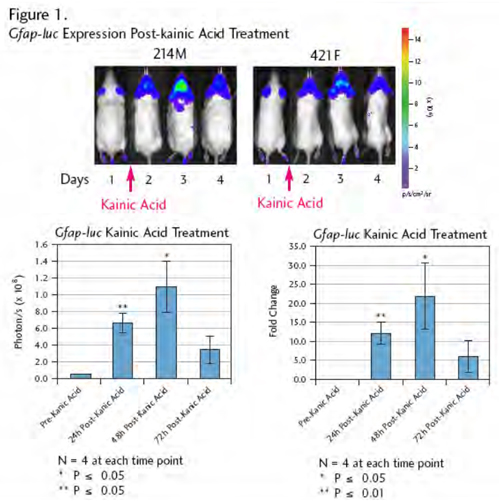
Figure 1: The Gfap-luc transgene was induced following kainic acid treatment. Age-matched adult mice were treated for four days. Treatment with kainic acid triggered higher luciferase expression in brain, with increased expression and neuronal damage to the hippocampus (data not shown).
Figure 1: The Gfap-luc transgene was induced following kainic acid treatment. Age-matched adult mice were treated for four days. Treatment with kainic acid triggered higher luciferase expression in brain, with increased expression and neuronal damage to the hippocampus (data not shown).
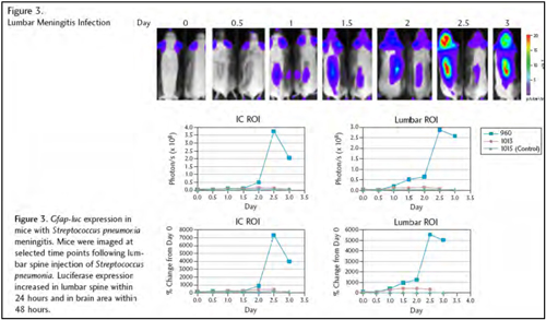
Meningitis infection study: The Gfap-luc transgene was induced to express luciferase in the brain (Figure 2) and the lumbar spine (Figure 3) of mice infected with Streptococcus pneumonia. 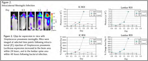
Imaging Recommendations
Anesthetize mice prior to injection of luciferin and measurement of luciferase transgene expression. Optimal imaging occurs between 10 and 20 minutes following intraperitoneal injection of luciferin.- Licensing
- Pricing - USD
- Pricing - EUR
- Pricing - DKK
- Select my Health Standard
- Get Custom Pricing Guide
Gfap-luc
Customers may purchase this model by signing the appropriate Terms of Sale (below) and providing a PO.
This model is sold under terms which grant perpetual use rights.
This model is sold under terms which grant perpetual use rights.
Pricing - USD
10501-EZcohort-4
| Item | Commercial | Nonprofit |
|---|---|---|
| Cryopreserved Model | US$11,500.00 | US$11,500.00 |
Cryopreserved models are invoiced upon shipment of recovered animals. Once orders are placed, the full purchase price will be applied if the order is canceled. For orders greater than 4 animals, please contact Taconic for options.
Fees for Taconic Transit Cages™ and freight are in addition to the price above.
Pricing - EUR
10501-EZcohort-4
| Item | Commercial | Nonprofit |
|---|---|---|
| Cryopreserved Model | 9.950,00 € | 9.950,00 € |
Cryopreserved models are invoiced upon shipment of recovered animals. Once orders are placed, the full purchase price will be applied if the order is canceled. For orders greater than 4 animals, please contact Taconic for options.
Fees for Taconic Transit Cages™ and freight are in addition to the price above.
Pricing - DKK
10501-EZcohort-4
| Item | Commercial | Nonprofit |
|---|---|---|
| Cryopreserved Model | kr.74.227,00 | kr.74.227,00 |
Cryopreserved models are invoiced upon shipment of recovered animals. Once orders are placed, the full purchase price will be applied if the order is canceled. For orders greater than 4 animals, please contact Taconic for options.
Fees for Taconic Transit Cages™ and freight are in addition to the price above.
Select my Health Standard
Need help choosing the right Taconic Biosciences health standard for your research?
Use the Health Standard Selector to enter your exclusion list. The tool will tell you which health standards meet your requirements.
Get custom pricing guide

Schedule A Scientific Consultation
Connect directly with a member of our Scientific Solutions team who can help you select the most appropriate model and maximize your experimental success.