Webinar Q&A — Histopathological Characterization of the Diet Induced NASH B6
Taconic Biosciences
Tuesday, September 1st, 2020
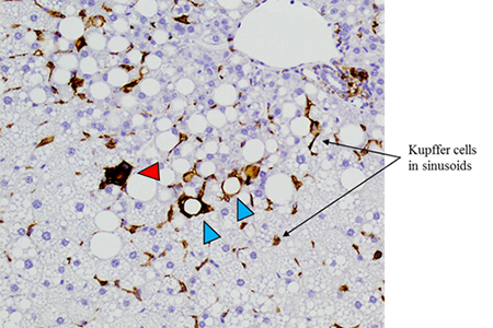
The Diet Induced NASH B6 displays hepatic crown-like structures, which are a unique histopathological characteristic seen in human livers with NASH. These hepatic crown-like structures are macrophages surrounding hepatocytes with large lipid vacuoles (blue arrows). Macrophages also form inflammatory cell aggregates with other leukocytes (red arrows). 20x magnification, F4/80 IHC on the liver from a C57BL/6NTac male mouse on NASH diet for 27 weeks.
Dr. Cynthia Besch-Williford of IDEXX BioAnalytics and Dr. Janell Richardson of Taconic Biosciences recently presented a webinar on histopathological characterization of the Diet Induced NASH B6. Histology remains the current gold standard for diagnosis of NASH in humans and it's an important readout for preclinical efficacy studies. Dr. Richardson provided a brief overview of NASH preclinical models and introduced the Diet Induced NASH B6. Dr. Besch-Williford, a veterinary pathologist board-certified in Laboratory Animal Medicine, reviewed quantitative and qualitative histopathological analysis of this model generated from mice on NASH diet for 27, 35 or 53 weeks along with age-matched C57BL/6NTac mice on a control diet.
Although the Q&A session went for 30 minutes, time constraints prevented discussion of all submitted questions. We present a full Q&A here.
The Diet Induced NASH B6 as a NASH model in the context of metabolic disease
Q: What do glucose and insulin levels look like in the Diet Induced NASH B6?
Dr. Janell Richardson: Insulin resistance is an important factor in the human metabolic NASH spectrum. Do we see it in our model? Historical data in the AMLN model indicates it is euglycemic, so blood glucose levels do not differ significantly from controls. We have also seen that in our Diet Induced NASH B6. We presented some data on this in our first NASH webinar. Blood glucose levels are not elevated compared to controls. However, HOMA-IR has been done historically in the AMLN model (original 301 AMLN diet) which show significantly elevated HOMA-IR levels compared to controls (at 30 weeks, Clapper et al. 2013). We are interested in looking at HOMA-IR in our model, but that data is not yet available. Some preliminary data suggests that our NASH B6 model is consistently hyperinsulinemic. Significantly elevated plasma insulin levels seem to be present, however, this still needs to be confirmed.
Q: Please clarify which B6 substrain is used for the Diet Induced NASH B6 and discuss what is known about the performance of B6/J versus B6/N substrains for NASH modeling.
JR: There are two major substrains of B6, the N (which stands for NIH) and the J (which stands for the Jackson Laboratory). These substrains arose before we understood genetic drift or how to prevent it. The data presented in this webinar is the B6NTac, the N substrain. So what are the differences between substrains and what does that mean for NASH phenotype? An anonymous pharmaceutical company looked at both substrains with AMLN-type diets, although the time course was different in terms of diet exposure, age and a few other things, but in general there were no large differences in the data. We did not review that data today, but some of it was presented in a previous webinar. What does this mean for day to day research? I can only speak to my own personal experience in the metabolic field. You can look at things like polymorphisms and there are genes such as the Nnt mutation which have been implicated in some of the differences between the J and N substrains. But what it often translates to is that I find the N substrain is just simply easier to work with. It typically has a narrowing of the standard error of the mean as it relates to things like bodyweight, blood glucose levels and insulin tolerance. Thus, it reduces the variance and provides a cushion for metabolic syndrome-dependent phenotype. I don't think there would be a dramatic difference in NASH phenotypes between the two models, but we don't have conclusive data on this point.
Q: Do you need to add another reagent to induce inflammation in addition to the NASH diet in C57BL/6NTac mice?
JR: No. All the data you've seen is purely just male C57BL/6NTac mice exposed starting at 6 weeks of age to the "310" diet and maintained on the diet for the period of time specified. They show inflammatory infiltration into their liver completely devoid of any other exogeneous administered toxins. Now can you use these mice and diet in combination with a toxin? Yes, but we have not explored what that might look like, for example with carbon tetrachloride, how that might influence the pathogenesis, whether you might be able to ask the same types of the questions. When you introduce a toxin, the mechanisms underlying that toxin's effect can certainly impact your ability to ask your question within the desired context.
Q: Are female mice suitable for NASH studies?
JR: C57BL/6 female mice (including C57BL/6NTac females) are resistant to a NASH phenotype. We do not know why. This is not the case in humans. To the best of my knowledge, there is no sex difference in prevalence within the human population. We do not offer female NASH mice for this reason.
Q: Have you looked at any potential cardiovascular and/or atherogenic effects such as aortic thickening or heart weight in this model?
JR: We haven't, and there's a good reason we haven't. There's a big dearth of rodent preclinical models which mimic what's seen in humans, for example vasculature changes or portal hypertension. If there is a model that recapitulates it within the spectrum of NASH, I am not aware of it. I know there are some that have some data that suggest positively on it. I doubt that our B6NTac is capable of recapitulating any of the mentioned indices.
Q: Is the "310" diet, sometimes called the "GAN" diet, available commercially?
JR: Yes, the "310" diet (D09100310) is a modified AMLN diet and retains much of the same properties that Amylin Pharmaceuticals described in their original publication on the use of this diet. The only difference is the modified part (source of the fat content) dealing with the substitution of palm oil for Primex. It is commercially available from Research Diets and that is the vendor that we use.
Q: Do you offer biochemical triglyceride analysis?
Dr. Cynthia Besch-Williford: Yes — IDEXX can measure serum lipids which are reflective of circulating changes in lipid composition. Liver triglyceride analysis isn't a standard offering, but can be performed. Given the special sample preparation needs, please contact the IDEXX BioAnalytics customer support services team for more details at 800-669-0825 or at idexxbioanalytics@idexx.com.
Liver Histopathology in NASH Mice
Q: In your opinion, what is the most specific NASH feature in the histopathology of the liver?
CBW: In terms of most important is the development of inflammation in response to lipid accumulation in hepatocytes. That is a key feature that is characteristic of NASH. The development of fibrosis occurs in consequence to inflammation. So the key part would be the presence of the inflammatory component, including the activated hepatic stellate cells, as well as the inflammatory cells that really generate the type of fibrogenic response that leads to that abnormal fibrosis.
Q: How homogenous are the various histopathological changes throughout the lobes of the liver? How can differences between lobes affect the ability to stage/score disease?
CBW: For this study, we looked only at one particular region of the median lobe of the liver. However, there have been studies that looked at interlobular disease manifestations and they tend to find that there is some variability, primarily in inflammation. But the degrees of lipid accumulation and fibrosis are fairly well represented across the different lobes of the liver. While there may be some influence of the heterogeneity of the inflammatory cell infiltrates, some of these other key features of NASH are preserved. So it does support the theory that a biopsy of a liver lobule may reflect fairly well the distribution of the changes across the entire liver.
Q: To what extent are hyperproliferative areas in the liver neoplastic versus regenerative?
CBW: Any type of proliferative change is on a scale from an early response that promotes hepatocyte regeneration to events that trigger uncontrolled proliferation which would be the definition of neoplasia. Some of the key features that are used to look at the difference between those two are morphological changes in liver architecture. When the architecture of the liver begins to be distorted by the hyperplastic response to produce new hepatocytes, that probably is one of the first changes that will prompt the concern that proliferation is uncontrolled. Another morphologic change as well is the de-differentiation of hepatocytes to a neoplastic phenotype. So when using morphologic changes alone, it's sometimes hard to make decisions as to whether a change is strictly hyperplastic versus neoplastic, but the key part in terms of the morphological appearance is whether or not that lobular architecture is entirely distorted so that the zones are no longer recognized.
Q: Is there any relation between the number of binucleated hepatocytes, mitotic figures and NASH?
CBW: That's a challenging question because mouse livers typically have binucleated hepatocytes, so for the most part that appearance is considered a normal feature. It does not necessarily indicate that there is an increased regeneration of cells or proliferation of hepatocytes that do not mature properly and retaining multiple nuclei. Mitotic activity can be found in normal livers to a small degree. In NASH livers, mitosis is not necessarily a feature associated with regeneration in hepatocytes. So I don't know that I have the knowledge of published work that correlates mitotic activity related to mitotic figures and NASH. Now there could be other markers of proliferation that might be more informative, such as Ki67 (nuclear proliferation marker).
Q: Does adipophilin stain all hepatic stellate cells?
CBW: In normal liver, the hepatic stellate cell when quiescent will contain a cytoplasmic lipid droplet, so it is expected that adipophilin would be a part of that cytoplasmic lipid-bound protein and would be detected by antibodies and IHC methodology. In the activated hepatic stellate cell, it's a little hard to gauge adipophilin labeling because those cells often are associated with lipid-filled hepatocytes and hepatic crown-like structures, so you already have adipophilin labeling of that large lipid-protein-bound membrane in those hepatocytes. So yes, it can be used to document the presence of hepatic stellate cells in the quiescent state. It's a little harder to know how to interpret the adipophilin labeling in the extremely steatotic liver because of the close association of those hepatic crown-like structures. Activated hepatic stellate cells also express smooth muscle actin or SMA, so can be visualized with antibodies to SMA using IHC methods.
Q: Do you have IHC to ubiquitin and keratin to better characterize the aggregates that were identified as Mallory-Denk bodies?
CBW: Our laboratory does not have those particular IHC assays. The literature has described use of IHC assays to target ubiquitin and cytokeratins because Mallory-Denk bodies are composed of ubiquinated proteins cytokeratin 8 and 18, and can be used to document the composition of the fibrils.
Q: How does histopathology in the NASH B6 model compare to alternative models such as ob/ob + stz?
CBW: Leptin-deficient (ob/ob) mice are hyperphagic and obese and develop hyperglycemia and hyperinsulinemia with insulin resistance. When fed a high-calorie diet, they develop marked hepatic steatosis and inflammation, but with minimal to low-grade fibrosis depending on type of high-calorie diet fed. Leptin plays a profibrogenic role in the liver in response to injury. Feeding a nutrient deficient diet, such as the methionine-choline deficient diets with elevated fat and sucrose/fructose content has been demonstrated to create hepatic fibrosis in this model.
Mice administered streptozotocin as neonates and fed a high-calorie diet develop steatosis, inflammation, fibrosis and hepatocellular carcinoma by 20 weeks, so the onset and progression of liver pathology is accelerated in streptozotocin-treated mice.
Q: Do these NASH B6 mice develop cirrhosis?
CBW: The Taconic NASH B6 mice at 53 weeks on diet had not developed cirrhosis. Livers from these mice had sinusoidal fibrosis that occasionally extended to bridge portal triads (bridging fibrosis). In cirrhosis, bridging fibrosis is more extensive, forming septae surrounding hepatic lobules and is accompanied by nodular hepatocellular regeneration.
Q: Do you have a service that can stain and score mouse liver histology slides that are provided by a customer?
CBW: We do. Depending on the goal of the study, we can use the same system as described here, the rodent NAFLD scoring system as well as using image analysis programs, to assess changes and quantify labeled or stained target structures. Generally, we want to ensure that the stain quality is such that it would allow our programs to work as we've designed them. So we would want to examine a photograph or a sample of a stained slide to assure that we can provide high quality service.
Q: Do you see recruited monocytes transformed to macrophages in the NASH model?
CBW: Studies to investigate macrophage phenotypes across the spectrum of disease development and to understand the role of resident versus recruited monocytes have been performed, but not as a component of this current study. This paper used Diet Induced Obese (DIO) B6 mice to examine resident versus recruited monocytes in NAFLD and the role of these phagocytic cells in disease development.
Q: Does microvesicular steatosis develop first and then and macrovesicular steatosis develop later?
CBW: We did not examine liver samples from Taconic B6 NASH mice on obesogenic diet prior to the 27 week time point. At this time point, both macro- and microvesicular hepatic steatosis were observed.
Use of Survival Liver Biopsies in NASH Mice
Q: What technique is used for survival liver biopsies in a mouse? Is it a needle aspirate?
CBW: Based on what I've read, these are not needle biopsies. It is more like a wedge biopsy in which a portion of the liver is removed by incision. After the sample has been collected, the margins that were sectioned in order to remove that piece are treated to minimize bleeding and then the lobe that was biopsied is returned into the abdomen and the incision is closed. The mice are often put on an antibiotic after surgery. It's more tissues compared to the size of the liver in a mouse than you would be able to get from a needle biopsy. So it gives you quite a bit of liver histology to review. There is a response in the liver to the biopsy in terms of healing, but it does allow you to opportunity to stage disease because you are looking at a much larger sample than would result from a core biopsy.
JR: My personal experience is using a wedge-based incision of the liver lobe and then you clot using a surgical sponge. Once clotted, you put the liver back into the animal.
Q: Are there any known impacts/influences of anesthesia for a biopsy on the future course of a NASH study?
JR: Survival-based liver biopsies in mice from start to finish should not take longer than about 10 minutes. We had a webinar on this recently. Add maybe 2 minutes for recovery, so 12 minutes from induction to recovery. It's really quite minimal. That doesn't mean it couldn't impact a study. The larger issue is always the post-operative care. That's my personal opinion from many surgical procedures in animals. It's about the post-operative care and observations to make sure that these animals recover quite quickly and maintain their normal food intake. Food intake in my opinion would be a much larger variable than anesthesia in NASH disease progression.
Model Selection
Q: Which model of NASH would be most appropriate when focusing on inflammation in the liver?
JR: For a model of inflammation in particular, what models would I be concentrating on? It's important to understand in the human condition that lipotoxicity is one of the hits, whether you are thinking two hit or the more modern multi-hit hypothesis. Metabolic syndrome is a chronic inflammatory disease. So when NASH goes bad, the pro-inflammatory cytokines are thought to reprogram the adult hepatocytes. According to a recent symposium, it kind of reprograms them to a fetal-like state. The important part is what starts all this. The thought is that it's metabolic syndrome and increased fat, and the problems start from there. So I would hone in on a model which has some characteristics of metabolic syndrome and move away from the toxin-based models. You can split inflammatory into two groups: liver and adipose. We don't have enough information on our model centering on adipose-based inflammation. We know that inflammation takes place in the liver. We have to do more exploration in quite a number of models. The benefit of NASH B6 in the context of that question is that's it simple, it's available and it's reliable. So could you choose a monogenetic model in combination with diet — does it show inflammation? Yes, but it's harder to get, you need more expertise to work with it because many of these models already are in a very heightened pro-inflammatory state, so it makes them incredibly sensitive to deal with. Picking a model where it's easy to be able to ask your question, it's able to at least recapitulate some of the mechanisms in the human inflammatory cascade and that you're able to assess the most basic question. In the context of leukocytes and macrophages, both of these are present in the NASH B6. When it comes to the cytokines, the immune cells, the supporting milieu, there are obviously differences between rodents and humans. I think there is more work to be done to really understand fine-tuned questions in the context of inflammation.
Improving Throughput for Histopathology Readouts in NASH Studies
Q: Histopathology is a complex technique which can have a more extended timeline for result reporting relative to other assays. For researchers attempting high-throughput screening in rodents, do you have any recommendations for ways to speed up the histopathology portions of studies?
CBW: Yes, I do. I think that we are in an era where artificial intelligence has really benefited the ability to train image analysis programs to view and score the changes in the liver. The value is these programs is sensitive measures of the degree of liver changes over time or with treatment. Being able to exploit these programs is a benefit. There are many on the market right now and they are generally validated by comparing the analysis to that of expert histologists or pathologists. They are recognized as valuable in sensitivity as well as specificity of lesion identification. These programs require quite a bit of training and consistency in histologic slide preparation and histochemical and immunohistochemical staining is needed to achieve reproducible results. Contract research organizations and pharmaceutical companies use image analysis to accelerate assessment of response of livers to treatment and to characterize disease models to complement traditional pathologist-generated data.
Taconic Biosciences' model generation team has produced about 5,000 models in the last 15 years, developing a globally-recognized reputation for advancing the work of in vivo researchers. Our scientific program managers are here to help you navigate the complexities of model generation.