Stephanie Oldham and Christian Rivera of AstraZeneca recently presented a
webinar on the use of a survivable liver biopsy in
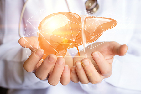
NASH mouse models, which is a technique that can provide valuable information on disease stage in individual mice prior to study initiation. In particular, it can be used to exclude outliers and properly randomize the remaining mice into study groups.
Due to time constraints, many of the questions attendees submitted during the Q&A session went unanswered. Here, we present the full Q&A.
Q: How much tissue is removed from the liver?
Stephanie Oldham and
Christian Rivera: In the mouse, we remove 30-50 mg of liver tissue.
Q: What is your recommendation for use of sterile surgical drapes when performing the liver biopsy?
SO &
CR: Any survival surgery requires a sterile field and aseptic technique. If you wish to use a surgical drape, it needs to be small. It can be difficult to expose left lateral lobe through a drape. You can certainly try it, and you may be successful with practice.
Q: What is the timing of analgesia delivery for the liver biopsy surgery?
SO &
CR: We administer analgesia via subcutaneous dosing up to an hour before the surgery so it's onboard during the surgery. After surgery, we administer topical analgesia directly to the sutures, before stapling. We also administer a second and third subcutaneous dose of analgesia 4-6 hours post-surgery and the next morning.
Q: What aftercare is recommended after the surgery?
SO &
CR: We maintain the mice in a warm cage for 30 minutes prior to replacing into the home cage. We also provide hydration gel in the home cage.
Q: High fat diet-induced NASH mouse models can be sensitive to stress. Do you observe any weight loss or phenotype regression after the liver biopsy?
SO &
CR: We do not. There is very little change in body weight after the surgery — they rebound incredibly quickly. They resume normal eating behaviors and physical activity within a day. We have not noticed any regression in phenotype.
Q: What mortality rate have you experienced with the liver biopsy?
SO &
CR: Our mortality rate is close to zero. Since the technique development phase, our mortality rate for actual studies is zero.
Q: What were some of the challenges you faced in developing this technique?
SO &
CR: The biggest challenge was exposing the left lateral lobe through the incision. We tried to minimize the length of that incision for healing purposes, but actually getting the liver out can be difficult. There is a lot of fat in these mice and sometimes fat will come out, which is not what you want. You must gently tuck the fat back in and try again. We use a cotton tipped applicator; gently place it under the left lateral liver lobe and use it to gently pull the lobe out through the incision. This avoids using forceps, which could damage the liver. Another method is to gently squeeze with your thumb and forefinger to "pop" the lobe out through the incision. Christian and I both prefer the cotton tipped applicator. Exposing the liver lobe is the most challenging part of the surgery. Taking the biopsy, applying the foam and suturing are all fairly simple.
Q: Does the gelatin foam efficiently control liver bleeding?
SO &
CR: Yes. Many times, there is very little bleeding when removing that wedge biopsy from the liver, particularly the more fibrotic the liver. The foam does absorb and stop bleeding. Rarely we see extra bleeding. If the foam piece is soaked with blood, remove it as it will not adhere to the liver. Just remove it and replace with a new piece of foam.
Q: Do you use a dry or saline-wetted cotton tipped applicator for the liver manipulation?
SO &
CR: We use it dry. You don't want to dry off the liver lobe, but with the applicator being dry it will help pull the liver lobe through the incision site more easily. A wet applicator is more slippery and won't provide the same leverage to pull the liver through. Once the liver is exposed, we lay it on a piece of gauze which has been wetted with sterile saline. This keeps the liver lobe hydrated.
Q: How long does it take per mouse to perform the biopsy?
SO &
CR: It takes 5-10 minutes per mouse. The longest part is often waiting for the mouse to be properly anesthetized.
Q: How long do you keep the mice after biopsy?
SO &
CR: We keep the mice for several months after the biopsy. There is a definite 3 week recovery time, and after that it depends on the duration of our treatment which is up to 12 weeks.
Application of the liver biopsy to other NASH rodent models
Q: Can this biopsy procedure be performed on mice which have been induced on a methionine choline deficient (MCD) diet?
SO &
CR: Procedurally you can perform this on any rodent, but with the MCD models there are logistical challenges. Prior to induction on an MCD diet, the mouse will have a healthy liver. After diet, the NASH phenotype and fibrosis progresses incredibly fast. Within just a few weeks you have a very fibrotic model. There isn't sufficient time to do a biopsy, recover and do any intervention or treatment before the end of the study because of the rapid onset of fibrosis with MCD diet. A biopsy prior to diet induction doesn't provide much information because it's a normal healthy liver.
Q: Do you have experience performing this type of liver biopsy in rats?
SO &
CR: We have not tried this in rats. There are reports that this procedure can be performed successfully in rats.
Histopathology analysis
Q: How do you identify inflammation by histology?
SO &
CR: We typically use Cd68 and Cd45 IHC staining for inflammation.
Q: What specific features are used to identify cell death and fibrosis?
SO &
CR: For cell death, you can look at macrophage staining and inflammatory staining, but we typically don't perform those. For fibrosis, we do collagen IHC, but you can use picrosirius red or galectin 3 or other markers indicative of collagen deposition.
Performing additional/terminal analyses
Q: If you want to perform additional analyses, can you take a larger portion of liver?
SO &
CR: Possibly. Particularly in ob/ob mice which have been conditioned on an AMLN diet, they are hyperphagic and they have very enlarged livers. Using C57BL/6 on an AMLN diet, the livers aren't as large. You must be careful not to remove too large a section of liver as it could jeopardize the function of the liver and the health of the mice throughout the study period.
Q: Is the left lateral lobe representative in terms of fibrosis which develops in other liver lobes?
SO &
CR: This is sort of a controversial. We always use the left lateral lobe and will continue to do so for comparison purposes. The medial lobe is commonly used by many other researchers. So the choice of lobe may be dictated by whether you want to compare your results with particular literature. I don't know whether the phenotype is consistent from lobe to lobe; it's not likely to be perfectly consistent. I would suggest picking a particular lobe and using it for all subsequent studies.
Q: Do you experience other differences between ob/ob and C57BL/6 mice when performing the liver biopsy?
SO &
CR: No. The biggest difference is just a somewhat larger and more fibrotic liver in the ob/ob mice as they develop fibrosis more quickly than the
C57BL/6. As the liver in ob/ob mice is more fibrotic and stiff, and we often see less bleeding in those mice. Ob/ob mice are reported to heal more slowly, but we have not observed that in our surgical model. We do not see any problems with healing and recovery in the ob/ob mice after liver biopsy.
Q: Is it possible to perform serial liver biopsies on the same animal?
SO &
CR: We have not tried this, but there are some reports that this can be done successfully. When we look at these mice 4-8 weeks later, you can see the liver heals incredibly well. You can see the foam piece and the structure of the liver is retained. If you take a subsequent biopsy from a different portion of the liver, this may be possible. However, undergoing a surgical procedure while on study might confound your results, so this is something to consider before performing a second biopsy.
Q: When performing a terminal analysis, from what portion of the liver is the sample taken? How does that terminal sample compare to the initial biopsy? Is the biopsy smaller, in a different region — does it affect your analysis in any way?
SO &
CR: That's a great question. We take the terminal sample from the same lobe (left lateral lobe). We take it adjacent to the initial biopsy, but not too close as we don't want to skew our data with inflammatory responses from the healing process. We perform all analyses using tissue from the left lateral lobe as this is the largest lobe and gives us consistency throughout the study. The terminal sample is larger so we can perform additional analyses. We typically use the biopsy only to assess collagen/fibrosis for randomization of study groups. The terminal sample is used for additional analyses such as steatosis, inflammation and biliary hyperplasia.
Translational aspects
Q: It was recently suggested that serum lipids correlate with NASH in patients. Do you see the same correlation in mice between serum lipids and histology?
SO &
CR: Yes, we do see elevated serum triglycerides in our NASH model and this correlates with liver lipids as well.
Q: In human patients, the biopsies are performed using needles. Can needle biopsies be performed in mice?
SO &
CR: I have not tried this, and we're not aware of anyone performing needle biopsies in mice. I am not sure exactly what syringes or needles are used for human biopsies. It might be difficult to hit the appropriate liver lobe without visualizing it.
 Watch the Taconic Biosciences' Webinars:

