Neurological disorders like Alzheimer's disease (AD) involve complex mechanisms that can be challenging to study. Taconic and LifeCanvas are collaborating to investigate how the disease develops and progresses, using advanced 3D histology with two Taconic mouse models used to study Alzheimer's disease.
Transgenic mouse models are crucial to understanding the mechanisms of neurological disorders like Alzheimer's disease and evaluating efficacious therapies. With those goals in mind, Taconic Biosciences has teamed up with LifeCanvas, an innovative provider of whole tissue processing and light sheet imaging, to characterize the distribution and density of beta-amyloid plaques and other pathological markers of Alzheimer's disease in two Taconic mouse models:
Additionally, IHC is only practical for analyzing a subset of tissue sections, which raises the risk of overlooking rare cell populations, disrupting neuronal connectivity, or inaccurately quantifying spatial relationships between certain cell types1. The latter is especially problematic in studying neurodegenerative disease, since spatial information is critical to assessing how microglia and other reactive glia interact with amyloid plaques.
The Taconic-LifeCanvas collaboration aimed to demonstrate proof of concept of 3D imaging in mouse models used to interrogate Alzheimer's disease, by characterizing 3D distribution and densities of beta-amyloid plaques and other pathological markers (including microglia and astrocytes) in brain tissue from Taconic ARTE10 and APPSWE mice.
The resulting 3D images and analyses revealed reactive glia, including microglia and astrocytes, clustering around and infiltrating amyloid plaques in the brain tissue of both ARTE10 and APPSWE mice. By using this novel imaging technology, the study produced more robust data, phenotyping, and molecular characterization of both Taconic AD models than would be possible with traditional IHC.
As investigators continue to study the formation of plaques as a marker of Alzheimer's disease, the combination of relevant disease-specific mouse models and advanced imaging technology can help them gain better insights into how the disease progresses and evaluate viable therapeutics.
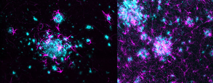
Left: Astrocytes marked by GFAP (magenta) cluster around β-amyloid plaques (cyan) in brain of 7-month-old homozygous ARTE10 (disease model #16347) male mouse from Taconic. SmartSPIM 15X objective.
Transgenic mouse models are crucial to understanding the mechanisms of neurological disorders like Alzheimer's disease and evaluating efficacious therapies. With those goals in mind, Taconic Biosciences has teamed up with LifeCanvas, an innovative provider of whole tissue processing and light sheet imaging, to characterize the distribution and density of beta-amyloid plaques and other pathological markers of Alzheimer's disease in two Taconic mouse models:
- The APPSWE, which expresses high concentrations of the mutant Aβ precursor protein carrying the Swedish mutation, and develops human AD-like amyloid pathology and displays memory deficits
- The ARTE10 (APP-PS1), which expresses high concentrations of the human Alzheimer β-amyloid (Aβ) precursor protein in addition to the human Presenilin 1 carrying the M146V mutation (PS1M146V) and develops human AD-like amyloid pathology and displays memory deficits
Using 3D Histology for a Better View
Assessing diseased brain tissue and the pathological markers associated with AD in mouse models requires high-resolution visualization of those markers, along with accurate localization and quantification of the associated neuroinflammation responses. Investigators traditionally have relied on 2D immunohistochemistry (IHC) to obtain these data; however, the approach is known to be inefficient, costly, and prone to errors. And while reconstructing 2D images in 3D improves the image resolution to some degree, the process of slicing samples can cause tissue damage.Additionally, IHC is only practical for analyzing a subset of tissue sections, which raises the risk of overlooking rare cell populations, disrupting neuronal connectivity, or inaccurately quantifying spatial relationships between certain cell types1. The latter is especially problematic in studying neurodegenerative disease, since spatial information is critical to assessing how microglia and other reactive glia interact with amyloid plaques.
The Taconic-LifeCanvas collaboration aimed to demonstrate proof of concept of 3D imaging in mouse models used to interrogate Alzheimer's disease, by characterizing 3D distribution and densities of beta-amyloid plaques and other pathological markers (including microglia and astrocytes) in brain tissue from Taconic ARTE10 and APPSWE mice.
Taconic Models Reveal Reactive Glia Clustering
Taconic sent whole brain samples from male and female ARTE10 and APPSWE mice to LifeCanvas, where they then conducted tissue delipidation, 3D immunolabeling with antibodies against beta-amyloid and glial markers IBA-1 and GFAP, imaging with a proprietary light sheet microscope, and image analysis. The study employed aged mice with the disease phenotype and compared disease pathology and progression across sex and at multiple time intervals, ranging from 8 weeks to 12 months.The resulting 3D images and analyses revealed reactive glia, including microglia and astrocytes, clustering around and infiltrating amyloid plaques in the brain tissue of both ARTE10 and APPSWE mice. By using this novel imaging technology, the study produced more robust data, phenotyping, and molecular characterization of both Taconic AD models than would be possible with traditional IHC.
As investigators continue to study the formation of plaques as a marker of Alzheimer's disease, the combination of relevant disease-specific mouse models and advanced imaging technology can help them gain better insights into how the disease progresses and evaluate viable therapeutics.