| Model No. | Nomenclature | Genotype |
|---|---|---|
| 10498 | FVB/NTac-Tg(Actb-luc)46Xen | Inquire for genotype |
β-actin-luc - Model 10498

View:
Select One
- Description
- Data
- Price & Licensing
- Overview
- Genetics
- Guides & Publications
- Applications & Therapeutic Areas
- Transit, Housing & Welfare
- Diet
Overview
Nomenclature: FVB/NTac-Tg(Actb-luc)46Xen
- Carries a 14 kb fragment of the murine β-actin promoter isolated from genomic DNA, a chimeric intron and modified firefly luciferase cDNA (Promega pGL3)
- Basal expression of the reporter is highest in skeletal muscle, thymus, skin, heart, bone, pancreas, and is detectable in all tissues, including white blood cells
- The reporter is constitutively expressed and is not significantly inducible
- Useful as a donor animal for studying the transplantation of various tissue types
Origin
The β-actin-luc mouse was developed by Caliper Life Sciences. The model was created by microinjecting a transgene containing a fragment of the murine β-actin promoter isolated from genomic DNA, a chimeric intron and modified firefly luciferase cDNA from pGL3. This transgene was microinjected into FVB/N zygotes. The resultant mice from founder line 46 were bred to FVB/NTac mice. Taconic received stock from Caliper in 2010. The line was embryo transfer derived. The line was maintained by mating wild mice and hemizygous mice.
This model is cryopreserved and available for recovery. Models can typically be recovered and delivered to customers within 12 weeks after order receipt. Purchase of this model includes perpetual use rights and a deliverable of four mutant animals at the Murine Pathogen Free™ health standard along with a genotyping protocol. For models which include a recombinase gene or multiple alleles, all alleles will be provided, but individual animals may not contain all mutant alleles.
Taconic’s Colony Management experts can design a plan to grow your colony faster.
Genetics
Guides & Publications
Initial Publication:
There is no specific publication describing the generation of these mice, but multiple publications exist demonstrating applications using the mice. See reference list.
Other publications:
- Cheeran MC, Mutnal MB, Hu S, Armien A, Lokensgard JR. (2009) Reduced lymphocyte infiltration during cytomegalovirus brain infection of interleukin-10-deficient mice. J Neurovirol. 15(4):334-42.
- Murphy CT, Moloney G, Macsharry J, Haynes A, Faivre E, Quinlan A, McLean PG, Lee K, O'Mahony L, Shanahan F, Melgar S, Nally K. (2010) Technical Advance: Function and efficacy of an {alpha}4-integrin antagonist using bioluminescence imaging to detect leukocyte trafficking in murine experimental colitis. J Leukoc Biol. 88(6):1271-8.
- Murphy CT, Moloney G, Hall LJ, Quinlan A, Faivre E, Casey P, Shanahan F, Melgar S, Nally K. (2010) Use of bioluminescence imaging to track neutrophil migration and its inhibition in experimental colitis. Clin Exp Immunol. 162(1):188-96.
- Schachtele SJ, Hu S, Little MR, Lokensgard JR. (2010) Herpes simplex virus induces neural oxidative damage via microglial cell Toll-like receptor-2. J Neuroinflammation. 7:35.
Applications & Therapeutic Areas
- Immunology
- Inflammation
- Reporter Lines
Transit, Housing & Welfare
Need more info? Click the live chat button or Contact Us
Diet
Data
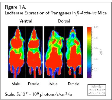
Figure 1A: Male and female mice were imaged in the ventral (left) and dorsal (right) regions. High basal luciferase expression was observed in males and females in feet, tail, ear, and mouth, all areas where bare skin is visible. Luciferase expression was observed in male gonads in some animals.
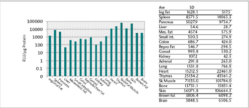
Figure 1B. β-actin-luc tissue expression survey from 2 male and 2 female mice (Fig. 1A). Data are expressed in Relative Light Units (RLU)/mg protein.
Tissue Expression of Luciferase signal
Luciferase expression of the β-actin-luc transgene was examined in extracts of tissue dissected from adult male and female mice from the β-actin-luc LPTA® animal model. Luciferase expression was highest in skeletal muscle, thymus skin, heart, bone, pancreas, and was detectable in all tissues examined including white blood cells (Fig. 1B). The bar graph is plotted with a logarithmic ordinate scale to graphically display all data. The expression data agree with ex vivo study data (not shown) and published work examining constitutive promoter-driven transgene expression.1,2 Luciferase expression was also examined in white blood cells from β-actin-luc mice (Fig. 1C (below)). Whole blood was harvested and spun to enrich for white blood cells. Cells were then serially diluted in a 96-well plate and placed in media containing luciferin substrate.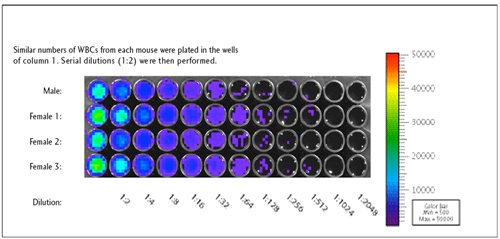
Figure 1C: Luciferase Expression in β-Actin-Luc Circulating White Blood Cells
Imaging Recommendations
Anesthetize mice prior to injection of luciferin and measurement of luciferase transgene expression. Optimal imaging occurs between 10 and 20 minutes following intraperitoneal injection of luciferin.- Licensing
- Pricing - USD
- Pricing - EUR
- Pricing - DKK
- Select my Health Standard
- Get Custom Pricing Guide
β-actin-luc - Model 10498
Customers may purchase this model by signing the appropriate Terms of Sale (below) and providing a PO.
This model is sold under terms which grant perpetual use rights.
This model is sold under terms which grant perpetual use rights.
Pricing - USD
10498-EZcohort-4
| Item | Commercial | Nonprofit |
|---|---|---|
| Cryopreserved Model | US$11,500.00 | US$11,500.00 |
Cryopreserved models are invoiced upon shipment of recovered animals. Once orders are placed, the full purchase price will be applied if the order is canceled. For orders greater than 4 animals, please contact Taconic for options.
Fees for Taconic Transit Cages™ and freight are in addition to the price above.
Pricing - EUR
10498-EZcohort-4
| Item | Commercial | Nonprofit |
|---|---|---|
| Cryopreserved Model | 9.950,00 € | 9.950,00 € |
Cryopreserved models are invoiced upon shipment of recovered animals. Once orders are placed, the full purchase price will be applied if the order is canceled. For orders greater than 4 animals, please contact Taconic for options.
Fees for Taconic Transit Cages™ and freight are in addition to the price above.
Pricing - DKK
10498-EZcohort-4
| Item | Commercial | Nonprofit |
|---|---|---|
| Cryopreserved Model | kr.74.227,00 | kr.74.227,00 |
Cryopreserved models are invoiced upon shipment of recovered animals. Once orders are placed, the full purchase price will be applied if the order is canceled. For orders greater than 4 animals, please contact Taconic for options.
Fees for Taconic Transit Cages™ and freight are in addition to the price above.
Select my Health Standard
Need help choosing the right Taconic Biosciences health standard for your research?
Use the Health Standard Selector to enter your exclusion list. The tool will tell you which health standards meet your requirements.
Get custom pricing guide

Schedule A Scientific Consultation
Connect directly with a member of our Scientific Solutions team who can help you select the most appropriate model and maximize your experimental success.