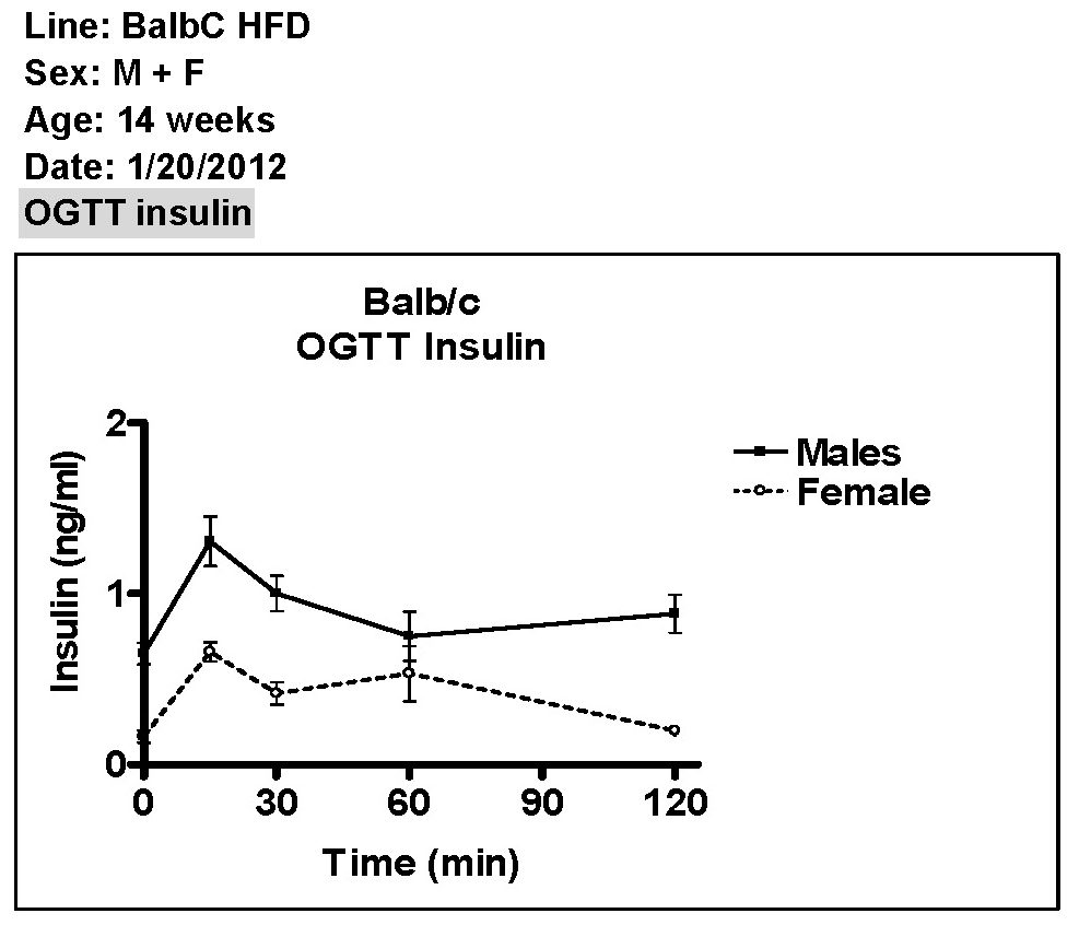
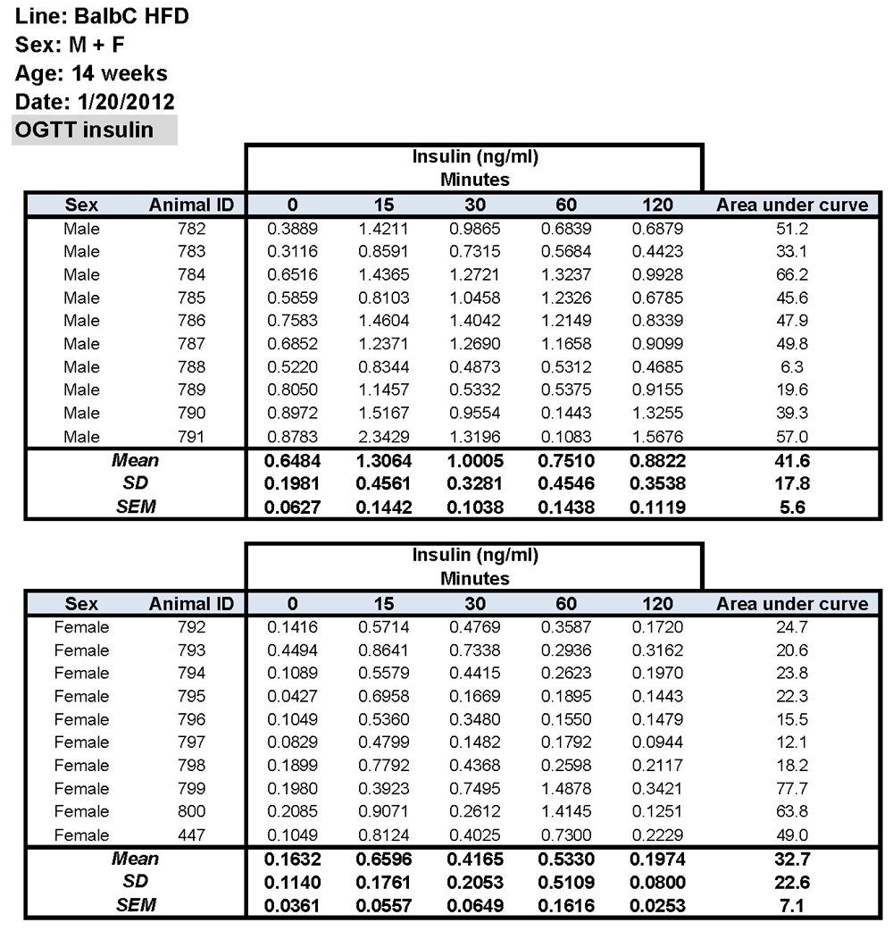
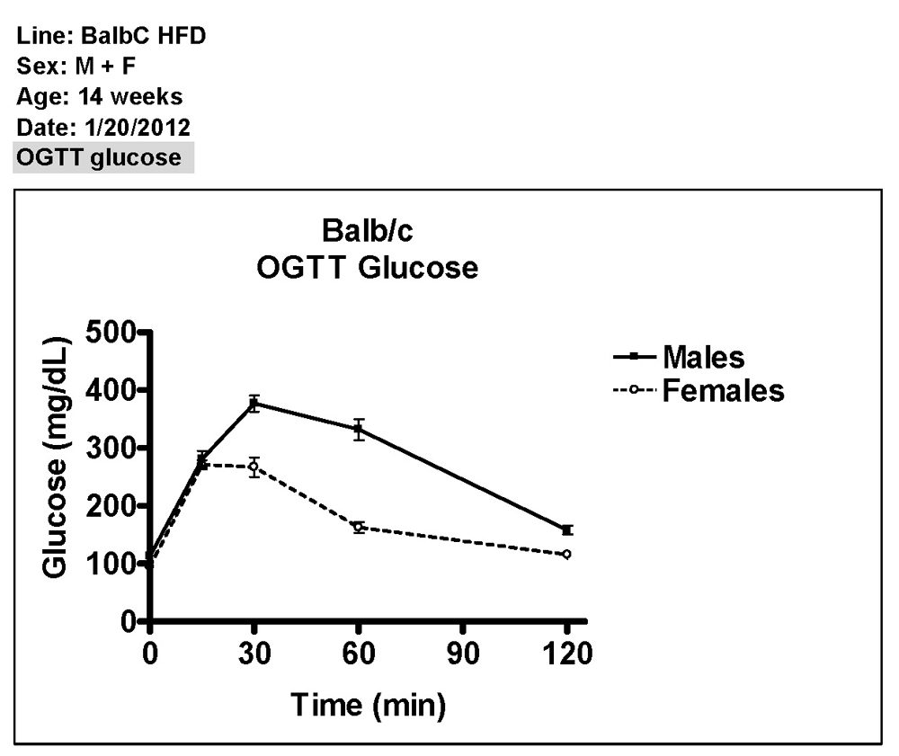
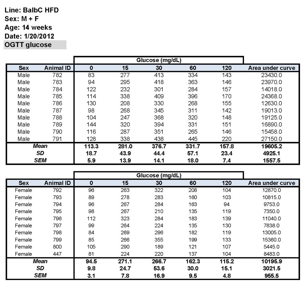
Oral Glucose Tolerance Test
Rationale:
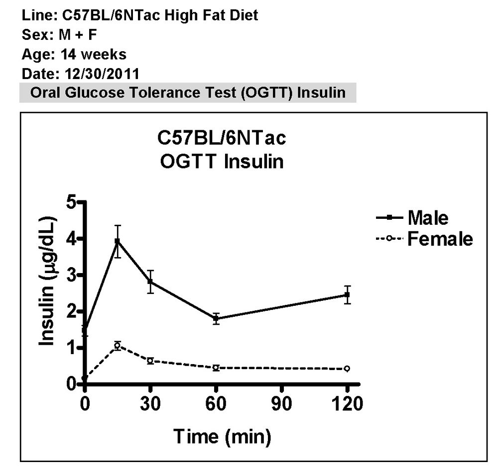
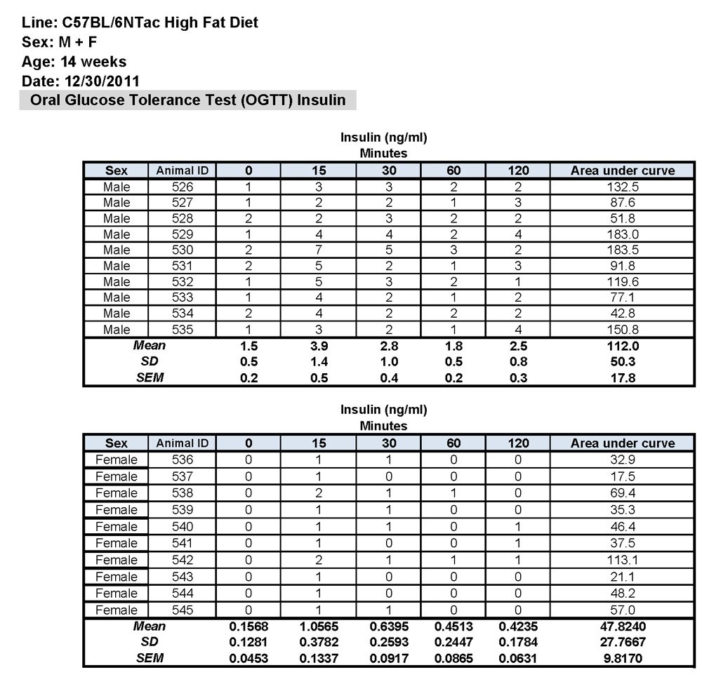
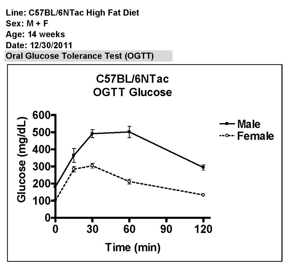
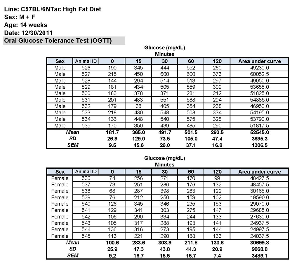
This assay is designed to identify genetically modified mice that exhibit alterations of metabolism associated with diabetes, obesity and cardiovascular disease. An oral glucose tolerance test (OGTT), in which the mice are challenged with a bolus of glucose and blood glucose and insulin levels are measured across a two hour time course is performed one week prior to the initiation of the HFD challenge. The mice are subjected to a seven-week HFD challenge using a Western diet. (Circulating levels of insulin, adiponectin, and cholesterol from samples taken before and after the HFD can also be measured, as well as the distribution of cholesterol in the VLDL, LDL, and HDL sub-fractions both pre and post the HFD challenge are analyzed using a polyacrylamide based system (Lipoprint).) The post HFD OGTT results are compared to those obtained before the start of the diet. The ability of genetically modified or pharmacologically treated mice to handle an oral glucose load, in combination with changes in insulin and adiponectin levels in response to the HFD, are assessed to identify genes or pharmacological agents affecting development of a diabetic or pre-diabetic state. Differences in cholesterol levels and cholesterol distribution are examined to establish if the genetic modification or the compound alters the response to the HFD.
Methods:
Prior to the test, the mice were fasted for 16 hours and transferred to a procedure room midway through the light phase of the Light:Dark cycle. Blood was obtained from a tail cut (by removing the distal 2 mm of the tail) and was assessed for baseline glucose levels using a One-touch Ultra 2 (Lifescan, Johnson & Johnson) glucometer. The remaining blood was processed for plasma that was later used to determine the fasting insulin levels. The mice then received 2 g/kg body weight of a 100 mg/ml glucose solution (Sigma, #G8769) in sterile water delivered by oral gavage. At 15, 30, 60, and 120 min after the administration of glucose, dried blood and tissue were quickly removed from the tail wound and blood was collected again to measure the glucose concentration and to prepare the plasma samples for measuring insulin levels. All of the plasma samples were frozen after collection and assayed later by electrochemiluminscence (MA2400 Mouse/Rat insulin kit K152BZC, Meso Scale Discovery) according to the manufacturer's recommendations.
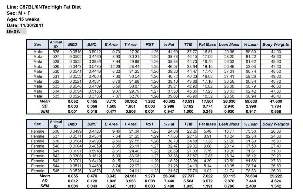
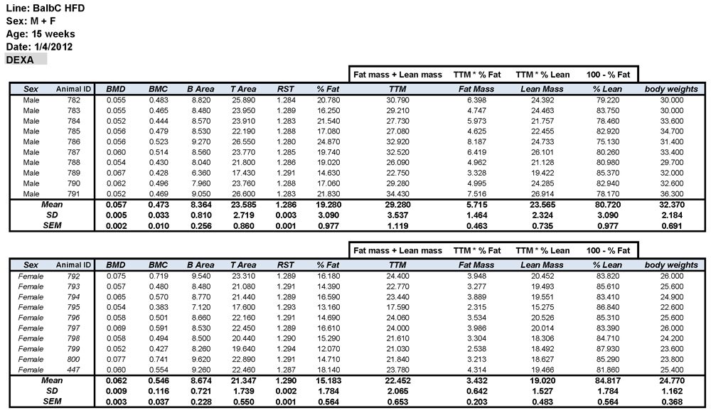
Body Composition by Dual-Energy X-ray Absorptiometry (DEXA)
The mice were anesthetized using 2.5% avertin (tert-amyl alcohol (Aldrich #15,256-3). Their body composition was assessed using the PIXImus2 X-ray unit (GE Lunar Corporation, Madison, WI) connected to a computer equipped with LUNAR PIXImus2 software. The head region of each mouse was excluded from the analysis. The following parameters were automatically measured or calculated by the PIXImus2 software: bone mineral density (BMD), bone mineral content (BMC), bone area (B Area), tissue area (T Area), percent fat, and total tissue mass (TTM). Fat mass, the percentage of lean mass, and lean mass, were calculated manually as follows: % Lean = 100 - % Fat
Lean Mass = TTM x (100 - % Fat)
Fat Mass = TTM x % Fat
Data Available by Strain:
C57BL/6NTac
BALB/cAnNTac
Welcome! Tell us a little about yourself